
Basic Airways - OPAs & NPAs
27th July 2021
Oropharyngeal airways ( also known as OPAs, “oral airways” or “Guedel airways”, named after the original designer Arthur Guedel (1)) and nasopharyngeal airways (also known as NPAs and nasal airways) are used to maintain an open airway.
Either device can be used depending on the indications for use and patient circumstances.
Oropharyngeal Airways
An unconscious patient is at risk of airway obstruction due to relaxed upper airway muscles - the pharyngeal dilator muscles (2) - or blockage of the airway by the tongue (3).

Due to the depth of an appropriately placed OPA, they can only be used in the unresponsive casualty patient who no longer has a gag reflex, to prevent vomiting of gastric contents.

Sizing the OPA
Although airways are simple to use, it is important to select an appropriate size: If the airway is too small, its distal end will be obstructed by the tongue, resulting in inadequate ventilation. If the OPA is too large, there is a risk of traumatic injury to the surrounding laryngeal structures (2), possible laryngospasm or can be obstructed by the epiglottis (3).
There are two common facial measurements recommended for determining the proper sized OPA: the distances between the maxillary incisors to the angle of the mandible, and the distance from the corner of the mouth to the angle of the mandible. A 2016 systematic review (4) determined the maxillary incisor to the angle of the mandible method to be most appropriate,
“In the maxillary incisors to the angle of the mandible group, the endoscopy did not identify any patient with complete obstruction of the airway by the tongue but in the corner of the mouth to the angle of the mandible group, 40% of patients had complete obstruction by the tongue.“

Kim JK, Kim SH, Min NH, Park WK. Determination of the appropriate sizes of oropharyngeal airways in adults: correlation with external facial measurements. European Journal of Anesthesiology. 2016;33:936-942.
For a rapid sizing, there is a strong correlation between casualty height and airway sizes. As such, for the average-sized casualty, the following OPA sizes can be used (5):
Average Height Male casualty - Size 9 OPA
Average Height Female casualty - Size 8 OPA
Inserting the OPA
Before inserting the airway, ensure the mouth is clear of secretions such as vomit, blood, or sputum.
Place the oral airway in the mouth with the concave side facing upwards towards the roof of the mouth.
As you are inserting the device, rotate the device 180 degrees into the correct position.
There is debate, without supportive evidence, to whether the flange should rest on the casualty’s lips or, on their teeth, under the lips. We would suggest the flange should rest where the OPA naturally sits.
Be sure to regularly assess the airway as, whilst the OPA keeps the airway open, it does not keep it clear from fluids.
OPA Contraindications
A responsive casualty with an intact gag reflex.
The casualty has a foreign body obstructing the airway.
Nasopharyngeal Airways
When using a nasopharyngeal airway, selecting the proper size is also important: If the NPA is too long, it will either enter the larynx and irritate the coughing and gag reflex, or be inserted into the vallecula, possibly causing an airway obstruction. If too short, the NPA will fail to separate the soft palate and tongue from the pharynx (5)

Sizing the NPA
The distal tip of the NPA is properly placed beyond the tongue base but should not be in contact with the epiglottis. It has been suggested that the ideal position for the NPA tip is 1cm above the epiglottis (6). The NPA’s length is a more important factor than its diameter in selecting the appropriate size of NPA (7).
There is NO correlation between the required size of the NPA and the casualty’s little finger, as is sometimes, erroneously, taught (8)
For rapid sizing, there is a strong correlation between casualty height and airway sizes. As such, for the average-sized casualty, the following NPA sizes can be used (9):
Average Height Male casualty - Size 7 NPA
Average Height Female casualty - Size 6 NPA
Optimal and rapid sizing of the NPA can be modified from these average sizes to take account of the subject’s height - the NPA should be measured from the nostril to the angle of the mandible. (10).
Inserting the NPA
The NPA should be lubricated with water-based lubricant.
With the fingers of the hand closest to the casualty’s head, resting on the casualty’s forehead, the thumb is used to lift the tip of the nose to open the nostrils.
The NPA is held in the other hand and positioned with the concave side facing down.
The tip is placed in the casualty’s nostril and the remainder of the NPA is positioned vertically.
The NPA is slowly inserted straight down - NOT following the angle of the nose.
If resistance is felt, the NPA should be withdrawn and insertion should be attempted in the other nostril.
The NPA should be inserted until the flange rests on the nostril.
If the casualty can tolerate one NPA, a second NPA should be attempted to double the cross-sectional area the casualty can breathe through.
Be sure to regularly assess the airway as, whilst the NPA keeps the airway open, it does not keep it clear from fluids.
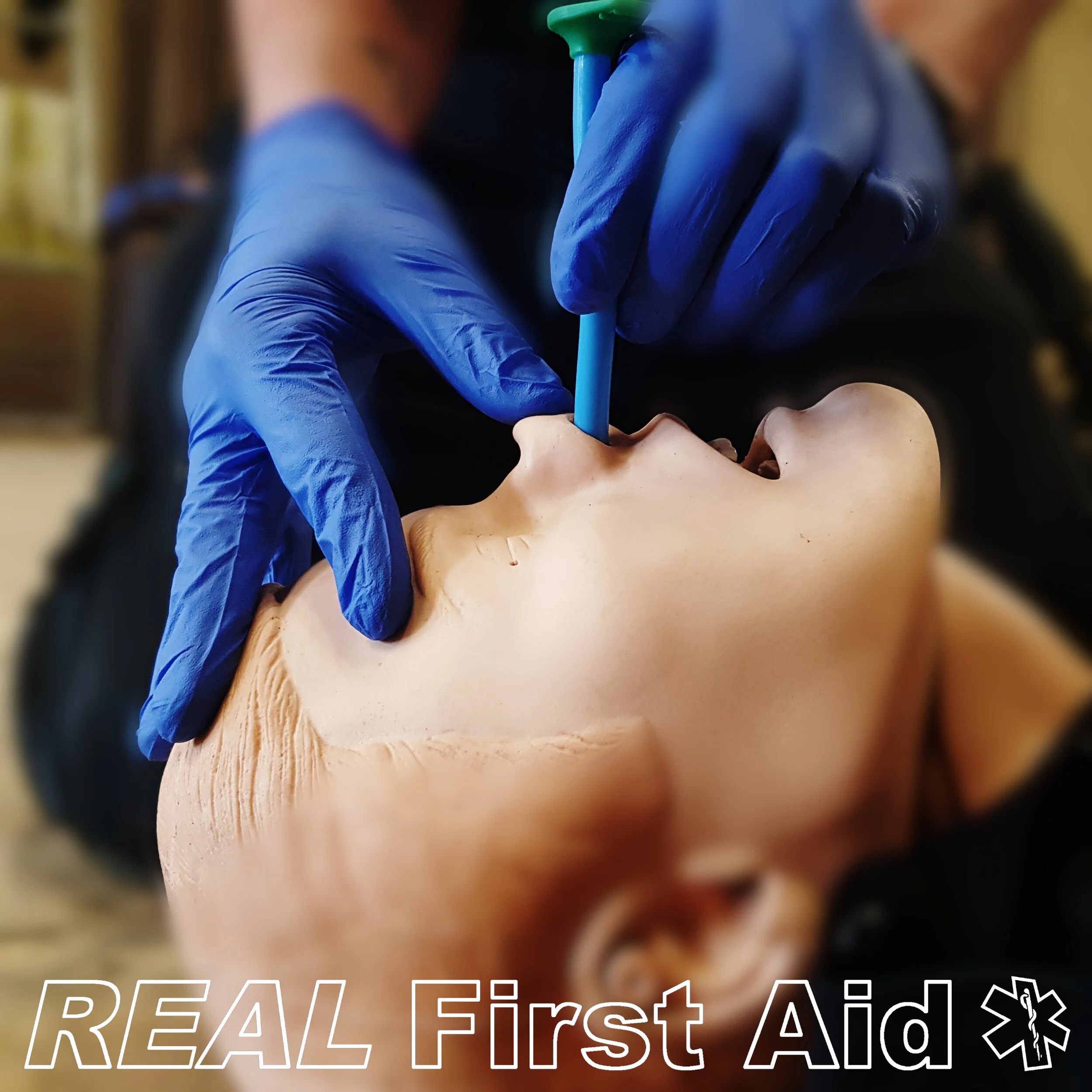
NPA insteted vertically.

NPA should NOT be inserted following the angle of the nose.
Intracranial placement
It is widely taught that a suspected or known basal skull fracture is a contraindication to NPA placement. This is based upon very few case reports (11, 12). This contraindication has been downgraded to a caution within JRCALC guidelines (13) and needs to be interpreted in the appropriate setting: faced with airway obstruction and the possibility of a basal skull fracture a responder must prioritise securing a patent airway prior to any further management.

Muzzi DA, Losasso TJ, Cucchiara RF. Complication from a nasopharyngeal airway in a patient with a basilar skull fracture. Anaesthesiology 1991;74:366–8.

Schade K, Borzotta A, Michaels A. Intracranial malposition of nasopharyngeal airway. J Trauma 2000;49(5):967–8.

Source: https://www.instagram.com/p/CPmOi8MLyWC/
In the absence of a CT scan the diagnosis of a basal skull fracture can only be presumed by the presence or absence of clinical features:
Blood from the ears or nose:
It must be appreciated that blood from the nose of patients with traumatic injuries is very common as is blood in the external ear (which has dripped into the ear rather than from within).
Cerebrospinal fluid from the ears or nose:
CSF is a clear fluid and its identification is difficult, even in hospital. The simplest tests, the halo sign or the presence of glucose, are easy to perform but have a low sensitivity and specificity (14, 15). Outside of a hospital and particularly in low light or wet prehospital conditions, the recognition of CSF from the nose or ears is near impossible.
Bruising around the mastoid process (Battle’s Sign) or both eyes (Racoon Eyes).
Common following trauma and is more likely to be associated with soft tissue injury than a basal skull fracture. Furthermore, the development of this bruising will take some time and is unlikely to have developed when the patient is in the early stages of resuscitation whether this is in the pre-hospital environment or upon arrival to the Emergency Department.
Therefore, the clinical features are vague and reliance upon their presence or absence may unnecessarily deter the use of the NPA. The evidence for avoiding NPAs in cases of basal skull fracture is based few case reports. Securing the airway in an emergency takes precedence over a suspected basal skull fracture (9).
In teaching the use of the NPA, the focus needs to shift from fear of contraindications to methods of safe placement to avoid intracranial placement. This needs to emphasise lifting the nostrils to reveal the nasal airway and the placement of the NPA vertically rather than along the angle of the nose, upwards towards the cribriform plate of the ethmoid bone.
NPA Contraindications
Known nasal airway blockages
Nasal fractures
Marked septal deviation
Coagulopathy (risk of epistaxis)
Obvious cerebrospinal fluid rhinorrhea
Known basilar skull fractures
The Silo or Rocket Technique
Key to airway patency is maximising airflow and reducing respiratory effort. A practice inserting two NAPs and one OPA (if tolerated) is known as the Silo or Rocket technique, with the aim of providing the maximum amount of cross-sectional area for the casualty to breathe through, reducing respiratory (or ventilatory) effort (16-18).

References
Guedel AE. (1933) Journal of the American Medical Association. 100:1862 (reprinted in “Classical File,” Survey of Anesthesiology. 10:515).
Kim HJ, Kim SH, Min JY, Park WK. (2017). “Determination of the appropriate oropharyngeal airway size in adults: Assessment using ventilation and an endoscopic view”. American Journal of Emergency Medicine. 35:1430-1434
Kim SH, Kim JE, Kim YH, Kang BC, Heo SB, Kim CK, Park WK. (2014) “An assessment of oropharyngeal airway position using a fiberoptic bronchoscope”. Anesthesia. 14;69:53-57.
Kim JK, Kim SH, Min NH, Park WK. (2016) “Determination of the appropriate sizes of oropharyngeal airways in adults: correlation with external facial measurements”. European Journal of Anesthesiology. 33:936-942.
Cherng CH, Huang G. (2013) “A modified lengthened nasopharyngeal airway”. Journal of Clinical Anesthesia. 25:240-246.
Stoneham MD. (1993) “The nasopharyngeal airway. Assessment of position by fiberoptic laryngoscopy”. Anesthesia. 48:575–80.
Roberts K, Whalley H, Bleetman A. (2005) “The nasopharyngeal airway: dispelling myths and establishing the facts”. Emergency Medical Journal. 22:394–6.
Roberts K, Porter K. (2003) “How do you size a nasopharyngeal airway?” Resuscitation.
56(1):19-23.
Roberts K, Whalley H, Bleetman A. (2005) “The nasopharyngeal airway: dispelling myths and establishing the facts”. Emergency Medical Journal. 22(6):394-6
Atanelov Z, Aina T, Amin B, et al. (2020) “Nasopharyngeal Airway”. [Updated 2020 Sep 25]. In: StatPearls [Internet]. Treasure Island (FL): StatPearls Publishing; 2021 Jan-. Available from: https://www.ncbi.nlm.nih.gov/books/NBK513220/
Muzzi DA, Losasso TJ, Cucchiara RF. (1991) “Complication from a nasopharyngeal airway in a patient with a basilar skull fracture”. Anaesthesiology. 74:366–8.
Schade K, Borzotta A, Michaels A. (2000) “Intracranial malposition of nasopharyngeal airway”. Journal of Trauma. 49(5):967–8.
Brown, S. N., Kumar, D. S., James, C., & Mark, J. (Eds.). (2019). JRCALC clinical guidelines 2019. Bridgwater: Class Professional.
Constantino PD, Janecka IP. (1998) “Cranial-base surgery”. In: Head and Neck Surgery—Otolaryngology, second edn. In: Byron J, ed. Bailey. LipincottRaven, Philadelphia, 1848–1853.
Chan DT, Poon WS, IP CP, et al. (2004) “How useful is glucose detection in diagnosing cerebrospinal fluid leak? The rational use of CT and Beta-2 transferrin assay in detection of cerebrospinal fluid fistula?” Asian Journal of Surgery. 27(1):39–42.
Weingart SD, Levitan RM. (2012) Preoxygenation and prevention of desaturation during emergency airway management.” Annals of Emergency Medicine. 59(3), 165–175.
Ley E, Webb T, Chesters A, Clarke B. (2015) "Essex and Herts Air Ambulance: a focused case series for pre-hospital practice". Journal of Paramedic Practice. 7:9, 438-445
Nutbeam T, Boylan M. (2013). The ABC of Pre-Hospital Emergency Medicine. Chichester, West Sussex, UK: John Wiley & Sons Ltd. p.23